
Imaging the mechanics and biology of joints,
and the implants that replace them
Our Research
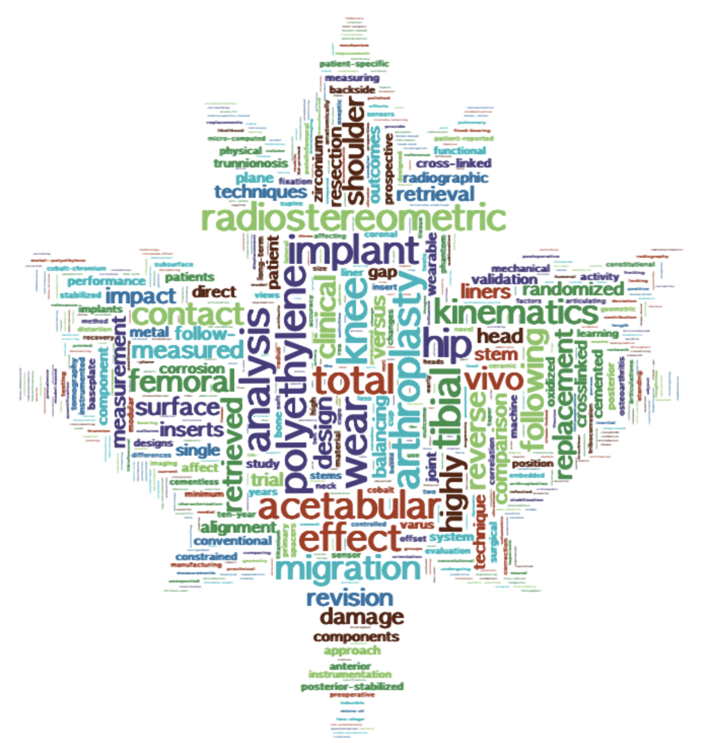
Joint replacement is one of medicine’s great successes, yet one in five knee replacement patients remains dissatisfied, and implants still loosen, wear, and become infected, often without early warning. The Teeter Lab develops imaging that reveals the mechanical and biological causes, from the arthritic joint before surgery to the implant decades after.
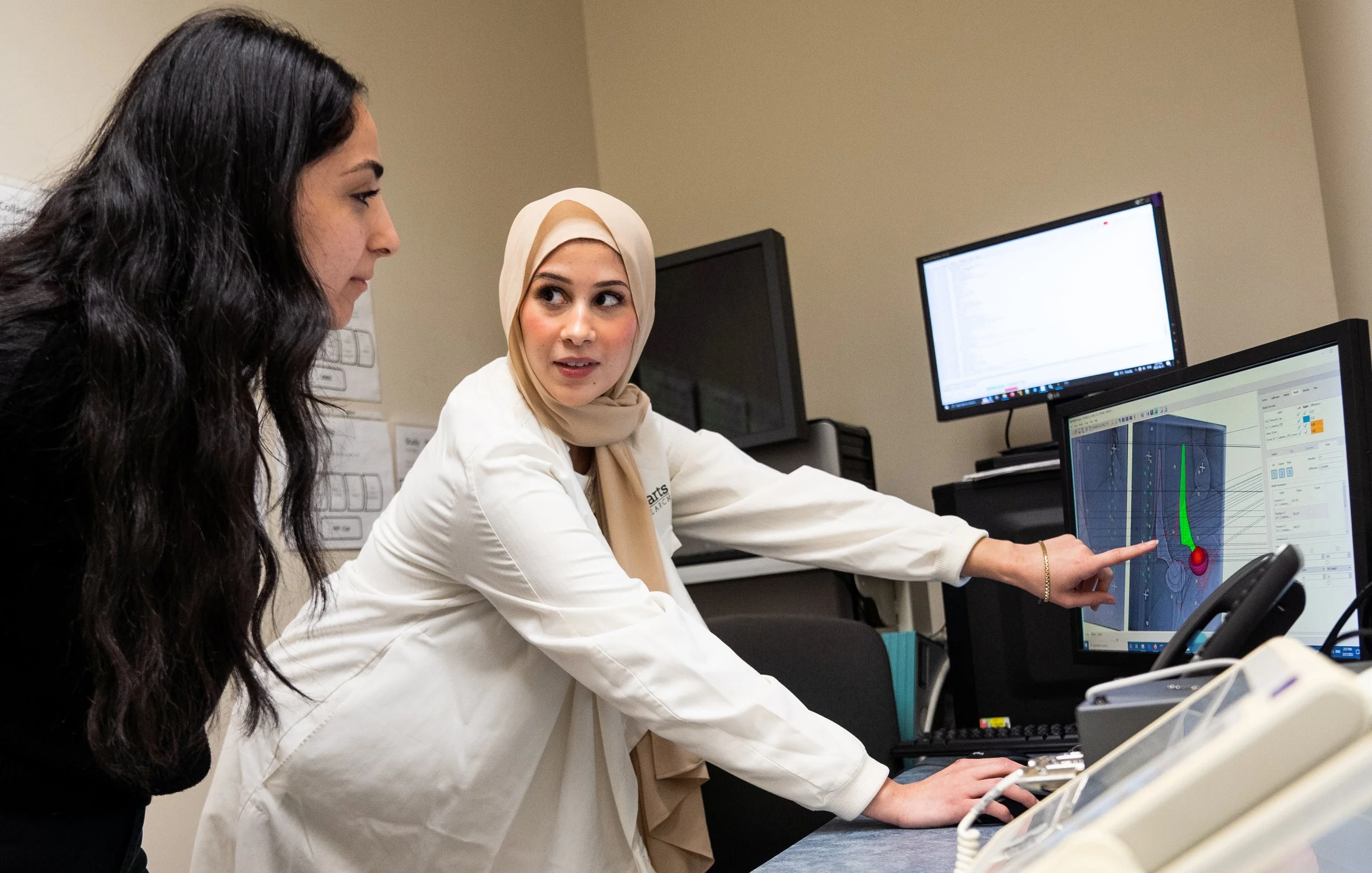
We create quantitative imaging tools and biomarkers that measure how joints and implants move, bond with bone, wear, and provoke inflammation. Our work spans RSA, CT, weight-bearing CT, MRI, and PET, linking what we see to how patients do, so surgeons can choose the right treatment for the right patient and detect problems before they fail.
With industry partners, we evaluate next-generation implants, surgical robotics, and biologic and digital technologies. We also build the enabling tools, including radiomics and machine-learning models, wearable and embedded sensors, and 3D-printed devices, that move imaging science into clinical practi
Check Google Scholar or follow Dr. Teeter on LinkedIn for the latest news. Contact us to explore collaborations in imaging, biomarker development, or technology translation.


Trainee Inquiries
Exceptional trainees interested in Medical Biophysics or Biomedical Engineering
may contact Dr. Teeter to discuss potential opportunities in the lab.